
Full text
Introduction
Hypertension in young adults was long regarded as a rare and largely functional condition; however, population data of recent decades have shown a steady increase in the prevalence of elevated BP among individuals aged 18–30 years and its association with cardiovascular events in middle age [1–3]. Organized contingents of security and defense agencies represent a distinct group: intense physical and psychoemotional loads, night duties, sleep deprivation and the use of energy drinks and nicotine create conditions under which transient BP elevations may consolidate into sustained hypertension [4, 5].
Departmental medical surveillance has traditionally relied on office BP measurement, which cannot distinguish white-coat, masked and sustained hypertension. Yet the masked form carries a prognosis comparable to that of sustained hypertension, and target-organ damage in the young develops subclinically [6, 7]. Pulse wave velocity (PWV), the cardio-ankle vascular index (CAVI), central hemodynamic parameters, markers of low-grade inflammation and oxidative stress, and albuminuria are considered mechanistic indicators of early vascular remodeling [8–11]; however, their combined assessment in young servicemen of the Republic of Uzbekistan has not previously been performed.
The aim of the study was to characterize the structure of BP phenotypes according to ambulatory monitoring, risk factors and early signs of target-organ damage, and to identify independent markers of vascular and myocardial remodeling in young men serving in the security and defense agencies of the Republic of Uzbekistan.
Materials and Methods
The study was performed at the Military Medical Academy within the framework of medical support for the security and defense agencies of the Republic of Uzbekistan. A total of 300 men aged 18–30 years on military service and subject to scheduled departmental medical examination were enrolled. The age structure was as follows: 18–20 years, 72 (24.0%); 21–23 years, 81 (27.0%); 24–26 years, 69 (23.0%); 27–30 years, 78 (26.0%). By unit profile, guard (118; 39.3%), training (96; 32.0%) and field (86; 28.7%) units were distinguished. Inclusion criteria were male sex, age 18–30 years, service in the security and defense agencies and the ability to complete the standard examination protocol; exclusion criteria were secondary hypertension and professional sports activity.
The design comprised two stages. At the first stage, clinical data, history, behavioral and metabolic risk factors, anthropometry, office BP, ECG and laboratory tests were recorded in all 300 subjects. At the second stage, a clinical-instrumental subgroup (n=147; 49.0%) was formed and underwent ABPM, echocardiography, treadmill testing, PWV and CAVI measurement, assessment of central hemodynamics and an extended biochemical profile. The remaining 153 subjects (51.0%) constituted the screening part of the study.
Office BP was measured by the auscultatory Korotkoff method on the right arm after 5 minutes of rest; the mean of the two closest readings was analyzed. ABPM was performed with a validated oscillometric recorder at 15-min intervals during the day and 30-min intervals at night; mean daytime, nighttime and 24-h SBP and DBP, pulse pressure, hypertension time index, magnitude and rate of the morning surge, short-term variability (thresholds: SBP >15 and DBP >14 mm Hg during the day, >12 mm Hg at night) and the nocturnal dipping pattern (dipper, non-dipper, over-dipper, night-peaker) were evaluated [13, 15]. The grade of hypertension was defined according to the current classification of BP levels for individuals over 18 years of age [3, 12]. Comparison of office BP (threshold 140/90 mm Hg) and ABPM allowed the phenotypes of sustained normotension, white-coat hypertension, masked hypertension and sustained hypertension to be distinguished. For comparative analysis, group I (true normotension: sustained normotension and white-coat hypertension, n=42) and group II (true hypertension: sustained and masked hypertension, n=105) were formed.
Echocardiography included calculation of left ventricular mass (LVM) and its index (LVMI), interventricular septal and posterior wall thickness, left atrial (LA) dimension and volume, end-diastolic and end-systolic volumes (EDV, ESV), stroke volume and ejection fraction (EF). Early myocardial remodeling was defined as increased LVMI and/or LA enlargement. Treadmill testing followed a stepwise protocol; a hypertensive BP response was recorded when SBP rose to 200 mm Hg or higher or when BP recovery was delayed. PWV was measured by the carotid-femoral and brachial-ankle methods [14], and CAVI with synchronous registration of pulse waves and ECG. Central hemodynamics were assessed by non-invasive reconstruction of aortic pressure: central systolic (cSBP) and pulse (cPP) pressure, the augmentation index normalized to a heart rate of 75 bpm (AIx@75), the peripheral-to-central SBP gradient (ΔSBP) and pulse pressure amplification (PPamp).
The laboratory panel included the lipid profile, glucose, creatinine with CKD-EPI estimation of GFR, microalbuminuria and the albumin-to-creatinine ratio (ACR), hs-CRP, IL-6 and TNF-α (enzyme immunoassay), NT-proBNP (immunochemiluminescent assay), malondialdehyde (MDA; thiobarbituric acid reaction), superoxide dismutase (SOD) activity and the integral oxidative stress index OSI = MDA/SOD.
Statistical processing was performed in IBM SPSS Statistics and Microsoft Excel. Quantitative data are presented as M±SD or Me [Q1; Q3]; groups were compared with Student's t-test and the Mann–Whitney test, and categorical variables with Pearson's χ² test with Yates' correction. Pearson and Spearman correlation analysis, odds ratios (OR) with 95% confidence intervals (CI), multiple linear and logistic regression and ROC analysis with determination of the AUC and Youden-index thresholds were used. Differences were considered significant at p<0.05.
Results
The clinical-instrumental subgroup comprised 147 servicemen (median age 21 [19; 22] years; height 180.13±7.05 cm; body weight 88.63±18.75 kg; BMI 27.27±5.24 kg/m2). Mean office BP in the subgroup was 144.22±16.23/89.83±8.26 mm Hg; 112 subjects (76.2%) had office BP ≥140/90 mm Hg. Comparison of office readings with ABPM showed that in 9 of the 35 subjects (25.7%) with normal office BP the ambulatory values corresponded to masked hypertension, whereas in 16 of the 112 (14.3%) with elevated office BP the 24-h values were within the normal range. The structure of BP phenotypes is shown in Figure 1.
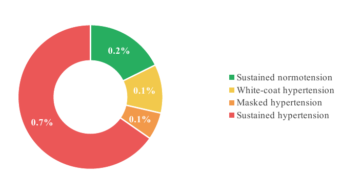
True hypertension (sustained and masked) was established in 105 subjects (71.4%; 95% CI 63.7–78.1) and true normotension in 42 (28.6%). Among the 96 subjects with sustained hypertension, 34 (35.4%) reported BP elevation lasting 6 years or longer, and only 3 (3.1%) monitored their BP on their own.
The groups did not differ in height (181.10±7.27 vs 180.22±7.11 cm; p=0.911), whereas body weight (91.31±16.21 vs 79.58±11.31 kg; p<0.05) and BMI (28.14±6.01 vs 24.54±5.29 kg/m2; p<0.05) were higher in group II. Overweight and obesity (BMI ≥25 kg/m2) were recorded in 73 (69.5%) vs 18 (42.8%) subjects (p=0.003), and obesity (BMI ≥30 kg/m2) in 42 (40.0%) vs 7 (16.6%) (p=0.008). The frequency of smoking (44.8% vs 35.7%; p=0.359) and of low physical activity (16.2% vs 11.9%; p=0.615) did not differ statistically. Total cholesterol (4.41±0.89 vs 4.08±0.69 mmol/L; p=0.039) and triglycerides (1.22±0.61 vs 0.94±0.55 mmol/L; p=0.013) were higher in group II at comparable glycemia. BMI correlated directly with 24-h SBP (ρ=0.258; p<0.001) and DBP (ρ=0.402; p<0.001). Three or more risk factors were present in 13 (12.4%) subjects of group II versus 1 (2.4%) in group I.
Office BP, ABPM, central hemodynamic and arterial stiffness parameters in the groups are given in Table 1.
| Parameter | Group I (n=42) | Group II (n=105) | p |
|---|---|---|---|
| Office SBP, mm Hg | 123.56±6.91 | 148.12±15.51 | <0.05 |
| Office DBP, mm Hg | 79.28±3.54 | 92.06±7.31 | <0.05 |
| 24-h SBP, mm Hg | 125.18±8.49 | 144.02±10.11 | <0.05 |
| 24-h DBP, mm Hg | 70.73±5.31 | 82.41±7.65 | <0.05 |
| Nighttime SBP, mm Hg | 119.58±10.44 | 135.86±11.74 | <0.05 |
| 24-h pulse pressure, mm Hg | 55.21±7.48 | 61.34±8.79 | <0.001 |
| Central SBP, mm Hg | 113.2±8.9 | 139.6±11.7 | <0.001 |
| Central pulse pressure, mm Hg | 39.4±6.5 | 53.7±8.6 | <0.001 |
| AIx@75, % | 6.1±7.1 | 15.4±8.4 | <0.001 |
| ΔSBP, mm Hg | 12.6±6.0 | 8.9±6.4 | 0.005 |
| PPamp | 1.41±0.18 | 1.27±0.17 | <0.001 |
| PWV, m/s | 6.45±0.74 | 7.68±0.92 | <0.001 |
| CAVI, units | 7.02±0.58 | 7.88±0.75 | <0.001 |
| PWV ≥7.5 m/s, n (%) | 6 (14.3) | 58 (55.2) | <0.001 |
High short-term SBP variability was recorded more often in group II during the day (53; 50.5% vs 7; 16.7%; p<0.001) and at night (46.7% vs 21.4%; p=0.005), as was high nocturnal DBP variability (37.1% vs 9.5%; p<0.001). A pronounced SBP load (time index >50%) was found in 99.0–100.0% of group II and in none of group I (p<0.001). The distribution of circadian profiles did not differ: the SBP non-dipper pattern was present in 45.2% and 48.6% (p=0.597), and a night-peaker profile was found in 2 subjects (1.9%) of group II. On treadmill testing (n=133), group II had higher SBP at rest (135.34±11.29 vs 123.94±8.76 mm Hg; p<0.008) and at peak exercise (170.38±14.97 vs 157.09±12.33 mm Hg; p<0.007); a hypertensive BP response was recorded in 86 (90.5%) vs 26 (68.4%) subjects (p=0.002) at comparable exercise capacity (9.86±2.27 vs 10.94±2.27 METs; p=0.182).
Renal function did not differ by creatinine (95.47±9.68 vs 95.34±12.06 μmol/L; p=0.872) or estimated GFR (98.09±12.21 vs 98.41±14.29 mL/min/1.73 m2; p=0.931), whereas microalbuminuria was higher in group II (0.072±0.11 vs 0.059±0.08 g/L; p=0.006). Echocardiography revealed greater LVM (158.4±17.21 vs 132.7±12.98 g; p=0.038), LVMI (99.02±19.11 vs 89.18±14.52 g/m2; p=0.031), LA volume (44.86±10.51 vs 41.47±12.09 mL; p=0.041), EDV (121.48±20.12 vs 111.14±21.73 mL; p=0.005), ESV (43.61±9.77 vs 38.32±10.19 mL; p=0.003) and stroke volume (79.27±5.44 vs 71.18±4.38 mL; p=0.003) in group II with preserved EF (64.71±3.02 vs 65.03±3.94%; p=0.236). Increased LVMI was found in 29 (27.6%) vs 4 (9.5%) subjects (p=0.018), LA enlargement in 26 (24.8%) vs 3 (7.1%) (p=0.011), and the combination of both signs in 19 (18.1%) vs 1 (2.4%) (p=0.006). The frequency of remodeling signs in the group II subgroups defined by the grade of hypertension (grade 1, n=41; grade 2, n=40; grade 3, n=7; isolated systolic hypertension (ISH), n=8; masked hypertension (MH), n=9) is shown in Figure 2.
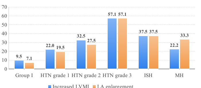
Remodeling signs were recorded both in masked hypertension (in more than 20% of cases) and in ISH, where LVMI reached 110.51±20.12 g/m2 (p=0.031 vs group I). Unfavorable features of the circadian profile were accompanied by a higher frequency of increased LVMI: 34% with high variability (p=0.012), 31% with nocturnal hypertension (p=0.018), 29% with a high BP load (p=0.021) and 27% with a morning surge at or above the threshold (p=0.033); the relationship, however, was not linear, since some subjects with a high BP load showed no signs of remodeling.
Analysis of central hemodynamics (Table 1) showed that cSBP in group II was 26.4 mm Hg higher, cPP was 14.3 mm Hg (36.3%) higher and AIx@75 was 2.5-fold higher. The peripheral-to-central SBP gradient decreased by almost one third and PPamp fell by 10%; reduced amplification (PPamp <1.25) was noted in 39 (37.1%) vs 5 (11.9%) subjects (p=0.002). PWV in group II was 1.23 m/s (≈19%) higher and CAVI 0.86 units higher; 55.2% of subjects with true hypertension exceeded the PWV threshold of 7.5 m/s. A stiffness gradient was observed across the group II subgroups: PWV was 7.29±0.79 m/s in grade 1, 7.74±0.84 in grade 2, 8.48±0.91 in grade 3, 8.11±0.83 in ISH and 7.06±0.72 m/s in MH; CAVI changed in the same direction (7.73, 7.96, 8.41, 8.20 and 7.57, respectively).
Markers of low-grade inflammation were significantly higher in group II: hs-CRP 1.7 [1.0; 2.9] vs 0.8 [0.4; 1.3] mg/L, IL-6 2.1 [1.5; 3.0] vs 1.2 [0.9; 1.7] pg/mL, TNF-α 3.3±1.0 vs 2.4±0.8 pg/mL (all p<0.001). With PWV ≥7.5 m/s compared with PWV <7.5 m/s, hs-CRP was 2.2±0.9 vs 1.1±0.6 mg/L, IL-6 2.8±0.7 vs 1.6±0.5 pg/mL and TNF-α 3.8±1.1 vs 2.7±0.8 pg/mL (all p<0.001). The median ACR in group II was twice as high (16 [9; 28] vs 8 [5; 13] mg/g; p<0.001), and ACR ≥30 mg/g was found in 19 (18.1%) vs 2 (4.8%) subjects (p=0.021). With increasing ACR (<10; 10–29; ≥30 mg/g), PWV (7.02±0.88; 7.63±0.91; 8.12±0.96 m/s) and cPP (48.5±8.2; 54.6±8.8; 59.3±9.4 mm Hg) rose stepwise. Oxidant status in group II was characterized by higher MDA (3.26±0.72 vs 2.43±0.61 μmol/L), lower SOD activity (1.47±0.27 vs 1.75±0.29 units) and a higher OSI (2.22±0.59 vs 1.39±0.44; all p<0.001); with PWV ≥7.5 m/s the OSI was 2.45±0.62 vs 1.58±0.48.
In multiple linear regression, the independent determinants of PWV were cPP (β=0.24; p=0.003), OSI (β=0.18; p=0.011), AIx@75 (β=0.16; p=0.031), hs-CRP (β=0.15; p=0.024) and ACR (β=0.13; p=0.042); the model explained 41% of PWV variance (R2=0.41; p<0.001). The results of logistic modeling of early myocardial remodeling are summarized in Table 2.
| Predictor | OR | 95% CI | p |
|---|---|---|---|
| Model 1: anthropometric and clinical-hemodynamic factors | |||
| BMI (per 1 kg/m2) | 1.12 | 1.05–1.21 | 0.002 |
| Length of service (per 1 year) | 1.19 | 1.07–1.34 | 0.001 |
| High BP variability | 1.87 | 1.02–3.41 | 0.041 |
| Nocturnal hypertension | 1.79 | 1.01–3.18 | 0.047 |
| Model 2: vascular and biochemical markers | |||
| PWV | 1.81 | 1.29–2.53 | <0.001 |
| AIx@75 | 1.22 | 1.04–1.40 | 0.013 |
| NT-proBNP | 1.19 | 1.06–1.34 | 0.004 |
| hs-CRP | 1.24 | 1.02–1.50 | 0.028 |
| ACR | 1.17 | 1.01–1.35 | 0.041 |
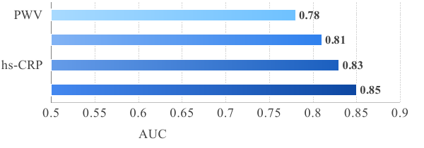
In ROC analysis, PWV had the highest discriminative ability for early remodeling (AUC=0.78; cut-off 7.55 m/s; sensitivity 74%, specificity 71%); the AUC was 0.71 for AIx@75 (cut-off 14%), 0.66 for hs-CRP (1.8 mg/L) and 0.65 for ACR (18 mg/g). Sequential inclusion of the markers in a combined model increased the AUC (Figure 3).
Discussion
The principal finding of this study is that, among young men referred for scheduled departmental examination, true hypertension was confirmed by the combination of office measurement and ABPM in 71.4%, while the phenotypes with discordant office and ambulatory pressure — masked and white-coat hypertension — together accounted for 17.0%. This figure is consistent with the reported prevalence of masked hypertension in population-based and clinical samples [6, 7] and confirms that a single office measurement at the unit medical post is an insufficient tool for risk stratification: in every fourth serviceman with normal office BP the ambulatory profile corresponded to hypertension, whereas in every seventh with elevated office BP it corresponded to normotension.
Importantly, signs of organ remodeling developed with preserved systolic function and were not rigidly linked to the severity of ambulatory indices: increased LVMI and LA enlargement were recorded both in masked hypertension and in ISH. The independent contribution of BMI and length of service to the probability of remodeling points to the cumulative effect of metabolic and service-related load, which agrees with data on the role of behavioral factors and the service regimen in the development of hypertension in conscripts and young servicemen [4, 5].
The data on central hemodynamics and arterial stiffness show that young men with true hypertension have already developed a pulse wave profile characteristic of a stiffer arterial bed: higher cPP and AIx@75, reduced amplification and higher PWV and CAVI. The prognostic value of PWV for cardiovascular events has been demonstrated in meta-analyses [8, 9], and the persistence of differences in CAVI, which is less dependent on the current BP level, indicates a structural component of stiffness. ISH deserves particular attention, as PWV in this subgroup approached the values of grade 3 hypertension: an isolated rise in the systolic component in the young should not be regarded as benign without assessment of arterial stiffness.
The coupling of inflammatory markers, oxidant imbalance and ACR with PWV and cPP fits the concept of mutual reinforcement of mechanical load and biochemical mechanisms of remodeling: subclinical inflammation precedes the development of hypertension [10], oxidative stress reduces nitric oxide bioavailability and accelerates extracellular matrix rearrangement [11], and increasing albuminuria reflects transmission of pulsatile energy into the microcirculation. The independence of each domain in the regression models and the increase in AUC with their sequential addition (0.78→0.85) justify the use of a combined assessment rather than a single index for early identification of the risk group.
The limitations of the study are its single-center cross-sectional design, the inclusion of men aged 18–30 years only, the relatively small size of the grade 3 hypertension, ISH and MH subgroups, and the absence of external validation of the models, which calls for verification of the obtained thresholds in independent samples and in prospective follow-up.
Conclusion
- In young men serving in the security and defense agencies, true hypertension according to office measurement and ABPM is detected in 71.4% of cases; masked and white-coat hypertension account for 6.1% and 10.9%, respectively, making ABPM a mandatory component of departmental screening.
- True hypertension is associated with overweight, atherogenic shifts of the lipid profile, high BP variability, a hypertensive response to exercise and early myocardial remodeling with preserved ejection fraction; the independent predictors of remodeling are BMI, length of service, high BP variability and nocturnal hypertension.
- The vascular phenotype of true hypertension in the young is characterized by elevated central pulse pressure, AIx@75, PWV and CAVI, reduced amplification, subclinical inflammation, oxidant imbalance and increasing albuminuria; arterial stiffness is independently determined by cPP, OSI, AIx@75, hs-CRP and ACR.
- PWV is the most informative single marker of early myocardial remodeling (AUC=0.78; cut-off 7.55 m/s); the combination of PWV, AIx@75, hs-CRP and ACR raises the AUC to 0.85 and can serve as the basis of a differentiated algorithm for early detection and follow-up of young servicemen at increased cardiovascular risk.