
Full text
Introduction
Apical periodontitis is a common consequence of root canal infection and represents the host defence response to microorganisms in the root canal system [1, 2]. It develops as a dynamic interaction between microbial factors and host defences, in which innate and adaptive immune cells, cytokines and bone-resorbing mediators determine the extent of periapical destruction [3–5].
IL-1 and IL-6 occupy a central place in this process. IL-1 is a potent bone-resorptive cytokine that induces osteoclast formation [6], and IL-6 participates in both the inflammatory and the regulatory phases of the lesion [7]. Chemokines and adhesion molecules control the recruitment of inflammatory cells into periapical tissue [8]. The response is not confined to the lesion: systematic reviews have linked apical periodontitis with higher circulating levels of inflammatory mediators and immunoglobulins [9, 10], and experimental lesions influence serum cytokine levels [11].
Most of this evidence comes from adults and animal models. Children differ from adults in the maturity of both innate and adaptive immunity [12], and secretory IgA, the principal effector of mucosal defence in the mouth [13], is among the measures that may vary with age. How these features shape the course of apical periodontitis in children and its response to treatment has been studied little. Adjunctive methods are also used: photodynamic therapy, for example, has been proposed for additional disinfection of the root canal system [14].
This study aimed to characterise systemic and local immune responses in children with different forms of apical periodontitis, to assess age-related differences and to compare immunological changes and recurrence during basic treatment and treatment supplemented with immunomodulators or with photodynamic therapy, probiotics and bacteriophages.
Materials and methods
Design and participants
This prospective cohort study with elements of controlled comparison was carried out over 18 months at the clinical base of a department of paediatric dentistry with the participation of a dental surgeon, an immunologist, a paediatrician and a medical statistician. It was approved by the local ethics committee (No. 04/D-21 of 12 March 2021) and followed the Declaration of Helsinki [15] and the principles of Good Clinical Practice; parents consented to their children’s participation. Reporting followed STROBE [16].
The study included 84 children aged 6–14 years who presented with apical periodontitis confirmed clinically and radiographically: acute serous (n=18), acute purulent (n=21), chronic granulating (n=23) and chronic granulomatous (n=22). Children were included if the disease was primary, with no interventions in the same area during the previous 12 months, no acute infectious disease during the previous 30 days and no history of immunosuppressive therapy. Exclusion criteria were immunodeficiency, allergic disease in the acute phase, active chronic infections including herpetic infection, severe somatic disease, oncohaematological disease and the use of antibiotics or immunostimulants within 3 weeks before observation.
Treatment
Group 1 (n=32) received basic therapy, which comprised mechanical and antiseptic treatment of the root canals together with antibacterial and anti-inflammatory drugs. Group 2 (n=27) additionally received immunomodulators selected on the basis of the preliminary immunogram from thymalin (5–10 mg intramuscularly every other day, 10-day course), polyoxidonium (6 mg rectally once daily at night for 10 days) and lycopid (1 mg orally twice daily for 10 days). Group 3 (n=25) additionally received photodynamic therapy, probiotics and oral bacteriophages (pyobacteriophage and staphylococcal bacteriophage). For photodynamic therapy, methylene blue was applied to the root canal walls and irradiated with a 660 nm laser for 1–2 min depending on canal volume, with control of temperature and exposure time. Probiotics containing Bifidobacterium lactis and Lactobacillus acidophilus were given as one capsule daily for 14 days from day 3 of treatment.
Clinical assessment
The examination included the history, clinical examination, thermometry, palpation and radiographic assessment of periapical structures. During treatment, pain, swelling, fever, lymph node status and blood counts were recorded daily. Clinical activity was assessed on an adapted scale of five items (pain on palpation, tooth mobility, fistula, soft tissue oedema and hyperthermia), each scored from 1 to 4, and the total score was compared before and after treatment. All children were followed for 3 months after treatment; recurrent inflammation of the tooth, a fistula or an abscess during this period was recorded as recurrence.
Laboratory and tissue analysis
Venous blood was drawn on an empty stomach between 8:30 and 9:00 into vacuum tubes without anticoagulant, delivered to a certified laboratory in thermal containers within 60 minutes and centrifuged; the serum was stored at −80 °C. Serum IgA, IgM and IgG were determined by enzyme-linked immunosorbent assay, circulating immune complexes (CIC) by polyethylene glycol precipitation, lymphocyte subsets (CD3+, CD4+, CD8+, CD16+/56+, CD19+) by flow cytometry with calibration of the cytometer before each series, and IL-1β, IL-6 and TNF-α by solid-phase enzyme-linked immunosorbent assay. Erythrocyte sedimentation rate and C-reactive protein were also recorded. Saliva was collected at a standardised time to measure secretory IgA (sIgA). Samples were analysed in duplicate by an operator unaware of group allocation. Laboratory measures were assessed before treatment and on days 7, 10 and 14.
Periapical biopsies obtained during abscess drainage or planned surgical intervention were fixed in 10% neutral formalin, embedded in paraffin and cut into 4–5 µm sections. Sections were stained with haematoxylin and eosin and immunohistochemically with polyclonal antibodies (Dako) to HLA-DR, ICAM-1, VCAM-1, TGF-β1, CD68 and CD3 with positive and negative controls. Infiltration was assessed morphometrically in five fields of view by two independent morphologists, who reached a consensus in doubtful cases.
Statistical analysis
Data were processed in Statistica 10.0 and R. Normality was checked with the Shapiro–Wilk test; two groups were compared with Student’s t-test or the Mann–Whitney test, and several groups with analysis of variance followed by Tukey’s test. Patients with different types of immune response were identified by k-means cluster analysis. Relationships between 27 clinical and laboratory variables and recurrence were assessed with Spearman’s correlation, and the association between immunomodulatory treatment and recurrence was estimated by multivariable regression and expressed as an odds ratio (OR) with 95% confidence interval (CI). Differences were considered significant at p<0.05.
Results
Systemic response by clinical form
Acute forms were accompanied by activation of the pro-inflammatory cytokine cascade, with elevated IL-1β, IL-6 and TNF-α. CIC levels in acute periodontitis were on average 63% above the age norm, with the highest values in purulent inflammation, and IL-6 concentrations exceeded the reference values 5.2-fold. On day 10, IL-6 in acute purulent periodontitis (87.4 pg/mL) was twice that in acute serous periodontitis (43.6 pg/mL) and 3.3 times that in chronic granulating periodontitis (26.2 pg/mL); in purulent inflammation, the CD4+/CD8+ ratio was the lowest (0.9) and CIC the highest (243 units/mL) (Table 1, Figure 1).
| Group | CD4+/CD8+ ratio | IL-6, pg/mL | CIC, units/mL | Salivary sIgA, mg/dL |
|---|---|---|---|---|
| Clinical form | Clinical form | Clinical form | Clinical form | Clinical form |
| Acute serous periodontitis | 1.3 | 43.6 | 179 | 126 |
| Acute purulent periodontitis | 0.9 | 87.4 | 243 | 92 |
| Chronic granulating periodontitis | 1.1 | 26.2 | 154 | 101 |
| Treatment | Treatment | Treatment | Treatment | Treatment |
| With immunomodulatory therapy | 1.6 | 18.7 | 93 | 158 |
| Without immunomodulatory therapy | 1.0 | 37.1 | 166 | 113 |
Values are group means. CIC, circulating immune complexes; sIgA, secretory immunoglobulin A.
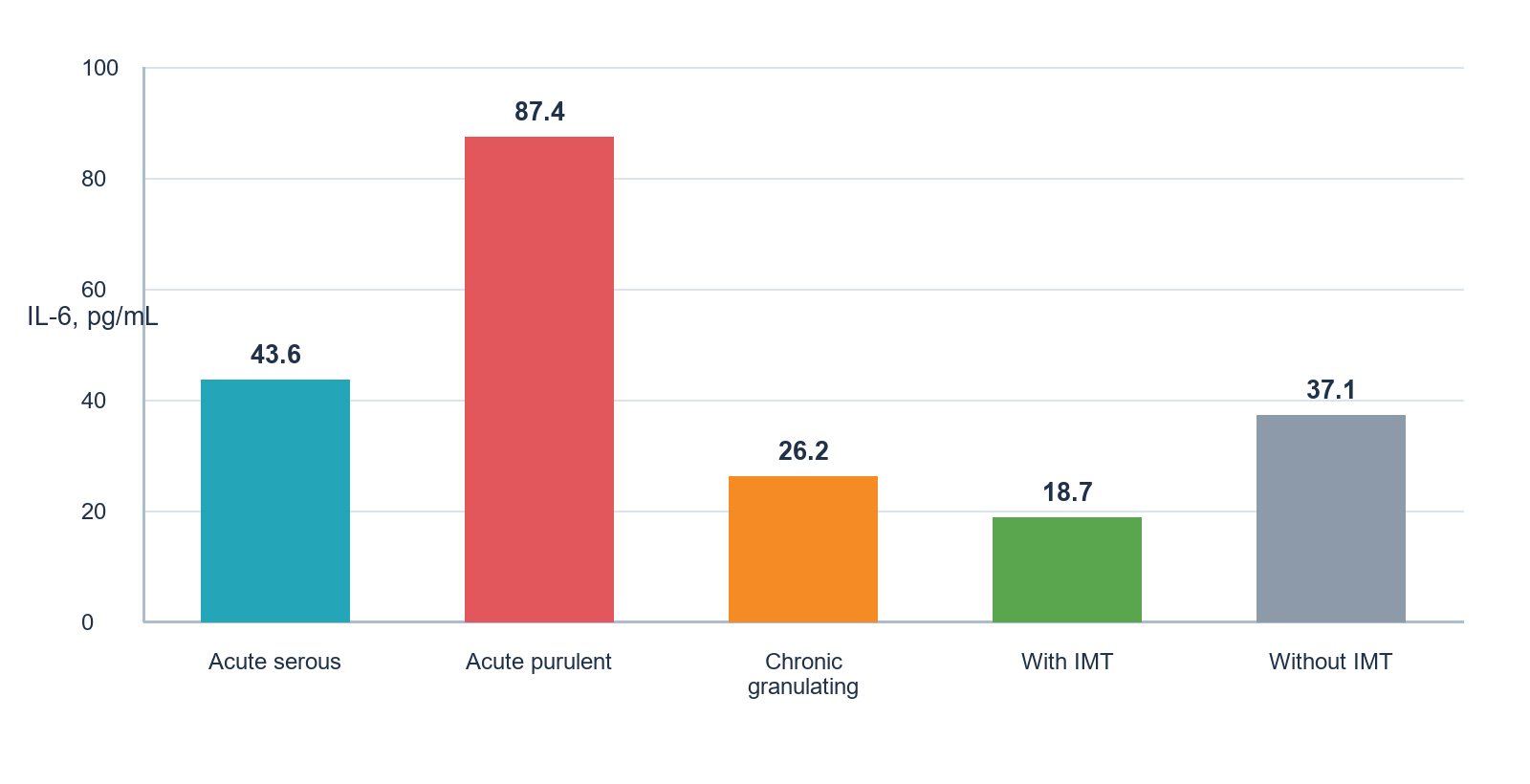
Group means. IMT, immunomodulatory therapy.
CD19+ B lymphocytes increased mainly in acute purulent periodontitis. In the chronic granulomatous form, CD8+ cells predominated over CD4+ cells, which accompanied a slowly progressing course and delayed tissue repair. In purulent periodontitis without fever, TNF-α was 28% higher than in children with a marked temperature reaction.
Local tissue response
Periapical biopsies showed high expression of the adhesion molecules ICAM-1 and VCAM-1, which persisted in the resolving phase of inflammation, and dense infiltration of granulation tissue by CD68+ macrophages. HLA-DR expression was high in the granulomatous form, indicating increased antigen-presenting activity.
Effect of immunotropic treatment
In children who received immunomodulators, the CD4+/CD8+ ratio normalised by day 10 and CIC by day 14. On day 10, compared with children treated without immunomodulators, their CD4+/CD8+ ratio was 60% higher, IL-6 and CIC were 50% and 44% lower, and salivary sIgA was 40% higher (Figure 2). Without immunocorrection, laboratory signs of inflammation persisted despite the regression of clinical symptoms; in some of these children, IL-6 decreased by day 7 but rose again by day 14.
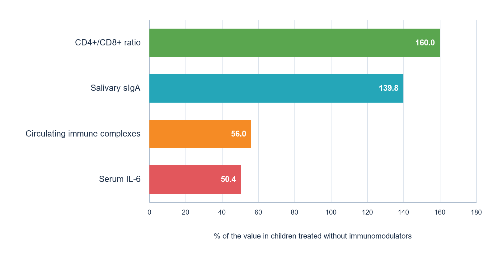
Values are expressed as a percentage of the corresponding mean in children treated without immunomodulators (100%); calculated from Table 1.
In children who received probiotics, salivary sIgA increased by an average of 51% from baseline by day 10. In multivariable analysis, the inclusion of immunomodulators in treatment was associated with a lower risk of recurrence (OR 0.43; 95% CI 0.29–0.66).
Age-related features
Immune measures changed with age (Table 2). The number of CD8+ T cells increased from 320 cells/µL at 6–8 years to 433 cells/µL at 13–14 years and salivary IgA from 91 to 124 mg/dL, whereas serum TNF-α was highest at 9–10 years (81.1 pg/mL). In the acute phase, CD8+ counts in children aged 6–8 years were on average 38% lower than in older children, particularly when treatment was given without immunocorrection.
| Age, years | CD8+ T cells, cells/µL | Serum TNF-α, pg/mL | Salivary IgA, mg/dL | Recurrence at 3 months, % |
|---|---|---|---|---|
| 6–8 | 320 | 74.5 | 91 | 28 |
| 9–10 | 367 | 81.1 | 103 | 21 |
| 11–12 | 415 | 76.3 | 118 | 14 |
| 13–14 | 433 | 69.8 | 124 | 11 |
Values are group means; recurrence is the proportion of children with recurrent inflammation during 3 months of follow-up.
The 3-month recurrence rate decreased from 28% in the youngest group to 11% at 13–14 years, in parallel with the increase in salivary IgA (Figure 3). In children with a recurrent course, salivary sIgA was on average 47% below the age norm, and 29% of them had CD19+ B-cell counts below 80 cells/µL.
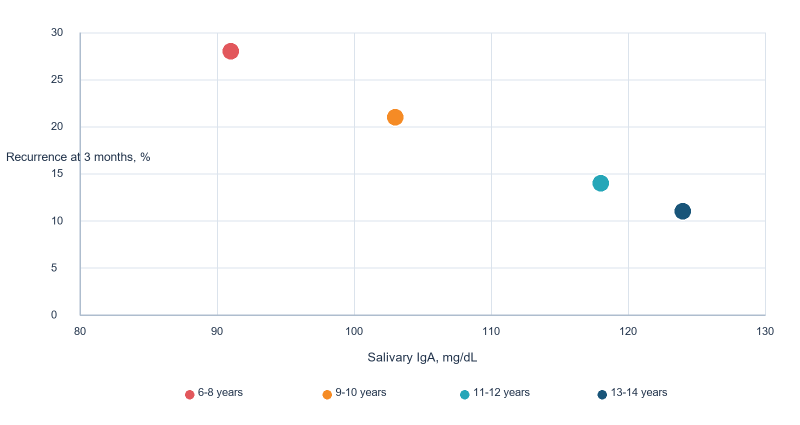
Each point represents the mean salivary IgA and the recurrence rate of one age group (Table 2).
Markers of severity and outcome
Correlation analysis of 27 clinical and laboratory variables identified IL-6, TNF-α, the CD4+/CD8+ ratio, CIC and salivary sIgA as the measures most closely related to the severity and outcome of inflammation.
Discussion
In this cohort, the immune response in apical periodontitis depended on the clinical form of the disease and on the age of the child. Acute purulent inflammation showed the most pronounced systemic changes, younger children had lower CD8+ counts and salivary IgA and more recurrences, and treatment supplemented with immunomodulators was associated with more complete normalisation of laboratory measures.
The increase in IL-6 and CIC in acute forms agrees with systematic reviews that found higher circulating inflammatory mediators and immunoglobulins in patients with apical periodontitis [9, 10] and with experimental evidence that periapical lesions influence serum cytokine levels [11]. IL-6 has both pro-inflammatory and regulatory functions in periapical lesions [7], which may explain why it reflected the activity of the process in our patients.
The persistence of ICAM-1 and VCAM-1 expression in the resolving phase is consistent with the role of adhesion molecules and chemokines in recruiting inflammatory cells to the periapical area [8]. Macrophage infiltration and high HLA-DR expression in chronic lesions correspond to the transition from innate to adaptive immunity and to the osteoimmunological mechanisms that sustain bone resorption [4–6].
The predominance of CD8+ cells in chronic granulomatous lesions and the low CD4+/CD8+ ratio in purulent inflammation are in line with the role of acquired immunity in the progression of chronic inflammatory lesions [17]. Lower CD8+ counts, lower salivary IgA and a higher recurrence rate in younger children reflect the gradual maturation of immune function during childhood [12] and the importance of secretory IgA for mucosal defence [13]. Young children with low salivary IgA may therefore be regarded as a group at increased risk of recurrence.
Immunomodulators were associated with better laboratory outcomes and a lower risk of recurrence. Because they were prescribed according to the immunogram rather than by random allocation, this association may partly reflect differences between the groups. Photodynamic therapy has been proposed as an adjunct for root canal disinfection [14]; in our cohort it was combined with probiotics and bacteriophages, so the contribution of each component cannot be separated. The increase in salivary sIgA during probiotic treatment requires confirmation in controlled studies.
Laboratory signs of inflammation persisted after clinical regression in children treated without immunocorrection. This supports immunological monitoring during treatment, particularly in young children and in recurrent disease. A minimal panel of serum IL-6, salivary IgA and basic lymphocyte subsets could be used for initial risk assessment.
Limitations
Treatment allocation was not randomised, and immunomodulators were selected according to the immunogram. The study had no healthy control group, so laboratory values were compared with age norms and reference values. The subgroups were small, the marker panel did not include IL-17 or IFN-γ, and recurrence was followed for 3 months only. The findings should be confirmed in multicentre randomised studies.
Conclusion
Apical periodontitis in children is accompanied by systemic and local immune changes that depend on the clinical form and on age. Acute purulent inflammation showed the highest IL-6 and CIC levels and the lowest CD4+/CD8+ ratio, and younger children had lower CD8+ counts and salivary IgA and a higher recurrence rate. Treatment supplemented with immunomodulators was associated with more complete normalisation of the CD4+/CD8+ ratio, IL-6, CIC and salivary sIgA and with a lower risk of recurrence (OR 0.43). Serum IL-6, salivary IgA and basic lymphocyte subsets may be used for risk assessment and treatment planning in children with apical periodontitis.