
Full text
Correspondence: Saidova Mukhlisa Akhrorovna, m.saidova@tashmeduni.uz, +998 91 443 82 93; Kamilov Khaydar Pozilovich, x.kamilov@tashmeduni.uz, +998 93 384 77 17.
Introduction
Subgingival instrumentation is the core of non-surgical periodontal therapy [1]. In a systematic review, it reduced probing depth by about 1.4 mm on average and closed most pockets within 6–8 months [2]. Residual deep pockets and bleeding after active treatment are, however, associated with further progression and tooth loss [3]. Maintaining the result is therefore as important as achieving it.
Patients with chronic digestive disease present a particular challenge. Their periodontal treatment takes place against a background of altered digestion, long-term medication and weakened local defence. Control of periodontal infection lowers serum inflammatory markers [4], and low-certainty evidence indicates that periodontal treatment combined with eradication therapy improves the outcome of gastric Helicobacter pylori treatment [5, 6].
Light-based adjuncts are used to modulate inflammation and support tissue repair. Meta-analyses of low-level laser therapy and other laser adjuncts report heterogeneous results and limited evidence of added clinical benefit [7–9], and European guidance does not support their routine use [1]. Photobiomodulation acts mainly through mitochondrial photoacceptors, and its most reproducible biological effect is a reduction in inflammation [10, 11]. The vacuum–laser technique combines irradiation with dosed negative pressure on the gingiva, which is intended to increase tissue blood filling during exposure; periodontal microcirculation can be monitored by laser Doppler flowmetry [12].
Nigella sativa seeds and their main active constituent, thymoquinone, have anti-inflammatory, antioxidant and immunomodulatory properties [13–16]. In patients with non-ulcer dyspepsia, N. sativa at 2 g daily achieved an H. pylori eradication rate that did not differ significantly from that of triple therapy [17]. Reviews describe possible benefits of N. sativa and thymoquinone in oral and periodontal disease [18, 19]; thymoquinone is active against oral bacteria in vitro [20], and a locally delivered thymoquinone gel improved clinical outcomes as an adjunct to scaling and root planing [21].
This study aimed to evaluate the clinical, laboratory, microbiological and microcirculatory outcomes of standard non-surgical treatment supplemented with vacuum–laser therapy and cold-pressed N. sativa oil in patients with gastrointestinal disorders and to assess the stability of remission over 6 months.
Materials and methods
Design and participants
This non-randomised controlled clinical study was carried out at the Tashkent State Dental Institute, and its results formed the basis of the institute's 2026 methodological recommendations on the combined treatment of inflammatory periodontal disease in gastrointestinal pathology. Reporting followed the TREND statement [22], and the intervention is described according to TIDieR [23].
The study included 200 patients aged 25–80 years with inflammatory periodontal disease. The main group comprised 107 patients with concomitant gastrointestinal disease: chronic gastritis (n=62), gastric or duodenal ulcer (n=9), colitis or enterocolitis (n=18) and gastro-oesophageal reflux disease (GORD; n=18). Their mean age was 51.7±12.4 years, 58 (54.2%) were women, and the gastrointestinal disease had lasted 7.8±3.6 years. The comparison group comprised 93 patients with periodontitis without gastrointestinal disease (50.3±11.8 years; 51 women, 54.8%). Values from 40 periodontally healthy individuals served as reference levels.
Allocation depended on gastrointestinal status, and the main group had a less favourable baseline profile. Severe periodontitis was present in 30 of 107 patients (28.0%) vs 15 of 93 (16.1%), and mild disease in 21 (19.6%) vs 34 (36.6%). Baseline BOP was 62.8% vs 45.2%, OHI-S 2.41 vs 1.92 points and probing pocket depth (PPD) 4.62 vs 3.71 mm. To take this imbalance into account, treatment effects were expressed as relative changes, and absolute values were compared at 3 months.
Interventions
Basic treatment was identical in both groups (Table 1). Scaling and curettage were performed by sextants according to pocket depth and tolerance, and root planing was carried out when subgingival deposits or root surface roughness were present. In patients with peptic ulcer disease or colitis, fewer procedures were performed per visit and the number of visits was increased. Antiseptic and anti-inflammatory therapy lasted 7–10 days and was followed by the first review.
| Component | Main group (n=107) | Comparison group (n=93) |
|---|---|---|
| Basic treatment | Professional oral hygiene; removal of supra- and subgingival deposits; scaling and curettage by sextants; root planing where indicated | Same |
| Antiseptic and anti-inflammatory therapy | 7–10 days, followed by the first review | Same |
| Oral hygiene training | Medium-bristle toothbrush and interdental brushes; technique checked at every visit | Same |
| Vacuum–laser therapy | Pulsed infrared laser, 890 nm, 80–100 Hz; negative pressure 0.04–0.06 MPa; 20–30 s per segment; 8–10 sessions every other day | Not used |
| N. sativa oil | First cold-pressed oil: gingival applications for 10–15 min twice daily and 5 mL orally twice daily 30 min before meals; 30 days | Not used |
| Follow-up | 7–10 days, 1, 3 and 6 months; repeat course if CCE for BOP is below 60% or pockets of 5 mm or more persist | 7–10 days, 1, 3 and 6 months |
CCE, coefficient of clinical efficiency; BOP, bleeding on probing.
Vacuum–laser therapy started after completion of basic treatment in the absence of acute inflammation and of exacerbation of the digestive disease. Vestibular and oral surfaces were treated in turn in each sextant; the actual number of sessions was 9.2±1.1. N. sativa oil was not given in cases of intolerance, exacerbation of the gastrointestinal disease or pregnancy, and in patients with peptic ulcer disease or colitis the regimen was agreed with a gastroenterologist before the course.
Outcomes
Complaints, OHI-S, PI, GBI, BOP, PPD and clinical attachment level (CAL) were recorded at baseline, 7–10 days after basic treatment and at 1 and 3 months. Laboratory follow-up included serum IL-1β, TNF-α and IL-6, salivary secretory IgA (sIgA) and PCR detection of Porphyromonas gingivalis, Tannerella forsythia, Treponema denticola, Prevotella intermedia, Fusobacterium nucleatum, Enterococcus faecalis and H. pylori. Gingival perfusion was measured by laser Doppler flowmetry as the index M in perfusion units (PU) [12]. At 6 months, the outcome was classified as remission, improvement without complete remission or no change, and recurrence of inflammation was recorded separately.
The coefficient of clinical efficiency was calculated as follows:
CCE = (I0 − I3) / I0 × 100%,
where I0 and I3 are the index values at baseline and at 3 months. A CCE of 60% or more for inflammatory indices corresponded to clinical remission. The protocol also set target values for OHI-S (CCE of at least 50%), PPD reduction (at least 1 mm), sIgA (at least 0.35 g/L), perfusion gain (at least 30%) and 6-month recurrence (below 15%). To describe stability, the proportion of the maximum improvement lost by 3 months was calculated as (I3 − Ibest) / (I0 − Ibest) × 100%, where Ibest is the best value during follow-up.
Statistical analysis
Data are presented as M±SD or n (%). Changes from baseline in the main group were assessed by Welch's t-test for continuous measures and by Fisher's exact test for proportions; because these tests ignore the within-patient correlation of repeated measurements, their p-values are conservative. At 3 months, the groups were compared on the primary indices, OHI-S and BOP, by Welch's t-test, and the difference was expressed as Hedges' g. Six-month outcomes were compared using relative risks (RRs) with 95% CIs on the logarithmic scale, risk differences with Newcombe CIs [24], the number needed to treat (NNT) [25], Fisher's exact test and the χ² test; proportions were given with Wilson CIs [26]. Because the groups differed at baseline, treatment effects were also expressed as relative changes, although this does not remove regression to the mean [27]. Sources of bias were considered using the ROBINS-I domains [28]. Associations in the main group were assessed by Spearman's rank correlation. Differences were considered significant at p<0.05.
Results
Course of gingival inflammation and oral hygiene
In the main group, OHI-S decreased from 2.41±0.34 to 0.86±0.21 points at 1 month and was 0.92±0.23 points at 3 months. PI changed from 2.18±0.31 to 0.72±0.19 and 0.78±0.22 points, GBI from 54.3±10.6% to 9.8±3.6% and 11.4±4.2%, and BOP from 62.8±11.3% to 12.6±4.1% and 14.8±4.8%. PPD decreased from 4.62±0.71 mm to 3.48±0.62 mm at 1 month and 3.26±0.58 mm at 3 months, a reduction of 1.36 mm, whereas CAL at 3 months was 91.4% of its baseline value. Changes in OHI-S, PI, GBI, BOP and PPD from baseline were significant at both time points (p<0.001).
The early response was similar in both groups (Figure 1). By 7–10 days, BOP had decreased by 65.9% in the main group and by 54.0% in the comparison group, and OHI-S by 53.5% and 43.8%, respectively. The curves diverged afterwards. In the comparison group, OHI-S rose from its best value of 1.08 points to 1.38±0.31 points and BOP from 18.4% to 26.7±8.4% by 3 months, so that 35.7% and 31.0% of the maximum improvement was lost. In the main group, the corresponding losses were 3.9% and 4.4%.
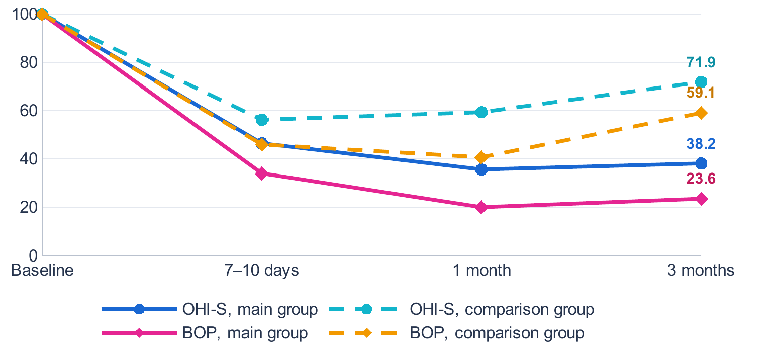
Group means are expressed as a percentage of the baseline mean; labels show the values at 3 months. Solid lines, main group; dashed lines, comparison group.
At 3 months, both indices were lower in the main group despite its higher baseline values: OHI-S was 0.92±0.23 vs 1.38±0.31 points (difference −0.46; 95% CI −0.54 to −0.38; g=−1.70), and BOP 14.8±4.8% vs 26.7±8.4% (difference −11.9 percentage points; 95% CI −13.9 to −9.9; g=−1.77); both p<0.001. CCE was higher in the main group for every index (Table 2). For the inflammatory indices, it exceeded the remission criterion of 60% in the main group (GBI 79.0%, BOP 76.4%) but not in the comparison group (41.6% and 40.9%). Relative to the comparison group, CCE for BOP was 86.8% higher.
| Index | Main group n=107 |
Comparison group n=93 |
Difference, pp | Target value |
|---|---|---|---|---|
| OHI-S | 61.8 | 28.1 | 33.7 | ≥50 |
| PI | 64.2 | 29.9 | 34.3 | – |
| GBI | 79.0 | 41.6 | 37.4 | ≥60 |
| BOP | 76.4 | 40.9 | 35.5 | ≥60 |
| PPD | 29.4 | 10.0 | 19.4 | – |
CCE = (I0 − I3) / I0 × 100%; pp, percentage points. Target values follow the protocol; for GBI, the remission criterion for inflammatory indices was applied. For PPD, the protocol target is a reduction of at least 1 mm, which was reached in the main group (1.36 mm).
Symptoms decreased in parallel. In the main group, gingival bleeding on brushing was reported by 86.0% of patients at baseline, 15.0% at 1 month and 18.7% at 3 months, and halitosis by 74.8%, 14.0% and 16.8%. Gingival oedema and hyperaemia were found in 91.6%, 12.1% and 14.0%, and burning or dryness of the mucosa in 57.9%, 19.6% and 17.8%. Each of these changes was significant at both time points (p<0.001). Dentine hypersensitivity decreased less (62.6%, 28.0% and 25.2%; p<0.001), and grade II–III tooth mobility changed little (29.9%, 26.2% and 25.2%; p>0.05).
Inflammatory, immune and perfusion measures
Serum cytokines fell markedly by 1 month and remained low at 3 months (p<0.001 vs baseline at both time points; Table 3). At 3 months, IL-1β, TNF-α and IL-6 were 2.6, 2.3 and 2.6 times lower than at baseline, corresponding to reductions of 61.6%, 57.0% and 61.2%, but they remained 33–45% above the reference values.
| Cytokine | Baseline | 1 month | 3 months | Reference level* |
|---|---|---|---|---|
| IL-1β | 14.6±3.2 | 5.1±1.6 | 5.6±1.8 | 4.2 |
| TNF-α | 17.2±3.8 | 6.8±2.0 | 7.4±2.2 | 5.1 |
| IL-6 | 12.9±3.0 | 4.6±1.4 | 5.0±1.6 | 3.6 |
Data are M±SD. *Mean values in 40 periodontally healthy individuals examined with the same protocol.
Salivary sIgA increased from 0.22±0.06 g/L to 0.39±0.08 g/L at 1 month and 0.37±0.08 g/L at 3 months (+68%), approaching the reference level of 0.42 g/L; in the comparison group, it changed only slightly. The perfusion index rose from 12.4±1.9 PU to 18.2±2.2 PU at 1 month and was 17.6±2.1 PU at 3 months; both measures differed from baseline at each time point (p<0.001). This gain of 41.9% exceeded the target of 30% and brought perfusion to 88.9% of the reference level (19.8 PU).
Microbiological changes
The detection of all seven microorganisms decreased by 3 months (p<0.001 for each; Figure 2). The largest absolute reductions concerned P. gingivalis, detected in 88 patients (82.2%) before treatment and in 28 (26.2%) at 3 months, and T. forsythia (80 and 24 patients; 74.8% and 22.4%). In relative terms, detection fell most for T. denticola (−70.8%) and least for H. pylori (−51.6%) and F. nucleatum (−59.3%). Oral H. pylori was found in 62 patients (57.9%) before treatment and in 30 (28.0%) at 3 months.
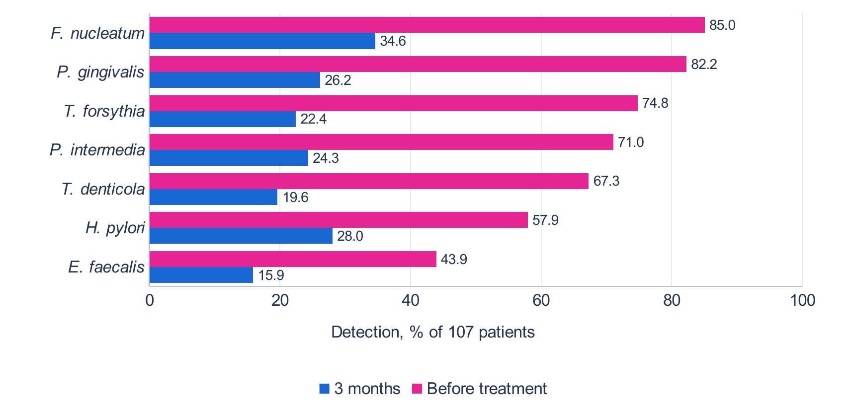
PCR detection; p<0.001 for each microorganism (baseline vs 3 months, Fisher's exact test).
Six-month outcomes
At 6 months, remission persisted in 77 of 107 patients in the main group (72.0%) and in 42 of 93 patients in the comparison group (45.2%). Improvement without complete remission was recorded in 24 (22.4%) and 31 (33.3%) patients, and no change in 6 (5.6%) and 20 (21.5%), respectively (χ²=17.8; p<0.001; Table 4). Recurrence occurred in 14 patients in the main group (13.1%; 95% CI 8.0–20.8) and in 29 in the comparison group (31.2%; 95% CI 22.7–41.2). The absolute risk reduction of 18.1 percentage points corresponds to an NNT of 6 (95% CI 4–16), and the main group met the protocol target of recurrence below 15%.
| Outcome | Main group n=107 |
Comparison group n=93 |
RR (95% CI) | Risk difference, pp (95% CI) | p | ||
|---|---|---|---|---|---|---|---|
| n | % | n | % | ||||
| Remission | 77 | 72.0 | 42 | 45.2 | 1.59 (1.24–2.05) | 26.8 (13.2 to 39.2) | <0.001 |
| Improvement | 24 | 22.4 | 31 | 33.3 | 0.67 (0.43–1.06) | −10.9 (−23.1 to 1.5) | 0.112 |
| No change | 6 | 5.6 | 20 | 21.5 | 0.26 (0.11–0.62) | −15.9 (−25.8 to −6.5) | 0.001 |
| Recurrence | 14 | 13.1 | 29 | 31.2 | 0.42 (0.24–0.75) | −18.1 (−29.3 to −6.6) | 0.003 |
RR, relative risk (main vs comparison group); pp, percentage points; p, Fisher's exact test. Remission, improvement and no change are mutually exclusive categories; recurrence was recorded separately.
The response depended on the gastrointestinal diagnosis (Figure 3). CCE for BOP was highest in GORD (84.2%) and chronic gastritis (77.5%), lower in colitis or enterocolitis (71.8%) and lowest in peptic ulcer disease (62.4%). Recurrence was recorded in 7 of 62 patients with gastritis (11.3%), 3 of 9 with peptic ulcer disease (33.3%), 3 of 18 with colitis or enterocolitis (16.7%) and 1 of 18 with GORD (5.6%). Remission at 6 months was maintained in 15 of 18 patients with GORD (83.3%), 47 of 62 with gastritis (75.8%), 11 of 18 with colitis or enterocolitis (61.1%) and 4 of 9 with peptic ulcer disease (44.4%). CCE in every subgroup of the main group exceeded that in the comparison group.
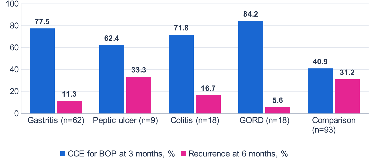
The comparison group had no gastrointestinal disease. Subgroups of 9–18 patients carry wide uncertainty. CCE, coefficient of clinical efficiency; GORD, gastro-oesophageal reflux disease.
Factors associated with the response
In the main group, CCE for BOP correlated directly with baseline sIgA (ρ=0.604) and with elimination of P. gingivalis (ρ=0.571), and inversely with gastrointestinal severity (ρ=−0.586). Recurrence was associated with baseline IL-1β (ρ=0.562) and with oral H. pylori (ρ=0.497). The number of vacuum–laser sessions correlated with the perfusion gain (ρ=0.538), baseline perfusion with PPD reduction (ρ=0.478), and the duration of gastrointestinal disease inversely with CCE for OHI-S (ρ=−0.512).
Discussion
In patients with gastrointestinal disease, standard non-surgical treatment supplemented with vacuum–laser therapy and N. sativa oil was followed by a durable reduction of gingival inflammation. The groups differed less in the speed of the initial response, which was similar at 7–10 days, than in the stability of the result. The comparison group lost about one-third of its improvement within 3 months, whereas the main group retained almost all of it, and recurrence at 6 months was less than half as frequent.
The reduction in probing depth of 1.36 mm at 3 months is close to the average effect of subgingival instrumentation reported in a systematic review [2]. The adjuncts therefore appear to have influenced the stability of inflammatory control more than pocket depth. This interpretation agrees with the small change in attachment level and with evidence that persistent bleeding and residual pockets determine long-term prognosis [3].
The contribution of the laser component has to be weighed against heterogeneous evidence. Meta-analyses of laser adjuncts to scaling and root planing have found small or inconsistent effects [7–9], and European guidance does not recommend their routine use [1]. Our protocol differed from most trials in combining the laser with negative pressure and in using a course of about nine sessions. The perfusion gain of 41.9% and its correlation with the number of sessions are consistent with the anti-inflammatory and vascular effects attributed to photobiomodulation [10, 11], but they do not prove a dose–response relationship.
The decrease in serum cytokines by more than half cannot be attributed to the adjuncts alone. Successful control of periodontal infection lowers systemic inflammatory markers by itself [4], and serial cytokine measurements were analysed in the main group only. The share attributable to N. sativa therefore remains uncertain. Its plausibility rests on the anti-inflammatory and immunomodulatory properties of thymoquinone [14–16] and on clinical data on locally delivered thymoquinone as an adjunct to scaling and root planing [19, 21]. The recovery of sIgA and its correlation with the response suggest that local defence is both a target of treatment and a predictor of its result.
The halving of oral H. pylori detection is clinically relevant because the mouth may act as a reservoir for reinfection. Periodontal treatment combined with eradication therapy has been associated with better gastric outcomes [5, 6], and N. sativa has shown anti-H. pylori activity in a clinical study [17] and antibacterial activity against oral bacteria [18, 20]. These results concern the oral detection of the bacterium.
The response depended on the digestive diagnosis. Patients with peptic ulcer disease had the lowest CCE and the highest recurrence, and gastrointestinal severity correlated inversely with the response. Together with the predictive value of baseline sIgA, this suggests that patients with severe digestive disease and low sIgA need closer maintenance and earlier decisions on repeat courses. In the treatment algorithm based on these data, the adjunctive block is intended for patients whose periodontal–gastric risk index, calculated from gastrointestinal severity, OHI-S, BOP and sIgA, exceeds 6.4 points. At 3 months, a CCE for BOP of 60% or more leads to maintenance care, 40–60% to a repeat course of the adjunctive block, and less than 40% to revision of the plan together with a gastroenterologist.
Limitations
The groups were not randomised and differed in gastrointestinal status and baseline severity, so confounding and regression to the mean may have affected the comparison [27, 28]. No group received standard treatment alone in the presence of gastrointestinal disease, and the separate effects of vacuum–laser therapy and N. sativa oil cannot be estimated. Serial laboratory and microbiological tests were analysed in the main group only. The peptic ulcer subgroup included nine patients, and follow-up was limited to 6 months.
Conclusion
In patients with inflammatory periodontal disease and gastrointestinal pathology, standard non-surgical treatment supplemented with vacuum–laser therapy and cold-pressed N. sativa oil was followed by a stable reduction of gingival inflammation (CCE for BOP 76.4% vs 40.9%), lower serum cytokines, higher salivary sIgA and gingival perfusion, less frequent oral H. pylori detection and fewer recurrences at 6 months (13.1% vs 31.2%). The response was weakest in peptic ulcer disease. A randomised trial in patients with gastrointestinal disease is needed to confirm the effect and to separate the contributions of the two adjuncts.